A Detail Of The Photomicrograph Shown In Fig 1b Where The Arrows Download Scientific
 Detail Of The Photomicrograph Shown In Fig. 1B, Where The Arrows... | Download Scientific ...")
(A) Detail Of The Photomicrograph Shown In Fig. 1B, Where The Arrows... | Download Scientific ...
(A) Detail Of The Photomicrograph Shown In Fig. 1B, Where The Arrows... | Download Scientific ... Download scientific diagram | (a) detail of the photomicrograph shown in fig. 1b, where the arrows indicate the locations of pb soap aggregates. Study with quizlet and memorize flashcards containing terms like outer fibrous layer, inner chondrogenic layer, lacunae and more.

Photomicrograph Illustrating | Download Scientific Diagram
Photomicrograph Illustrating | Download Scientific Diagram Film is a stern judge of how good the microscopy has been prior to capturing the image. it is essential that the microscope be configured using köhler illumination, and that the field and condenser diaphragms are adjusted correctly and the condenser height is optimized. A student carried out two experiments to investigate the progress of the reaction shown in fig. 6.1. potato tissue was used as the source of the enzyme. six pieces of potato were cut, each measuring 20 mm × 10 mm × 10 mm. Examine the photomicrograph on the right, taken using an oil immersion objective, the highest practical magnification. your solution’s ready to go! our expert help has broken down your problem into an easy to learn solution you can count on. This chapter gives you the background for making excellent photomicrographs. it is divided into two parts. the first part deals with digital cameras and digital micrographs. the second part deals with film cameras and film micrographs.

Photomicrograph Diagram | Quizlet
Photomicrograph Diagram | Quizlet Examine the photomicrograph on the right, taken using an oil immersion objective, the highest practical magnification. your solution’s ready to go! our expert help has broken down your problem into an easy to learn solution you can count on. This chapter gives you the background for making excellent photomicrographs. it is divided into two parts. the first part deals with digital cameras and digital micrographs. the second part deals with film cameras and film micrographs. Many fundamental topics are discussed in detail in this article including the reciprocity law, characteristic curves, exposure bracketing, exposure calculations, and filter factors. The degree of detail that can be seen in an image is known as the resolution. the tinier the individual points of information on an image – for example, the pixels on a monitor – the better the resolution. to see the very smallest objects, you need a microscope with very high resolution. The resolution of a microscope is a measure of the smallest detail of the object that can be observed. resolution is expressed in linear units, usually micrometres (μm). Photomicrography – the process of taking photographs with the microscope – used to be a highly specialised skill taking many years of practice to achieve good results. digital cameras have largely changed that, as the instant viewing of results allows errors to be corrected immediately.

Solved 4. Examine The Photomicrograph In Fig. G-53. Describe | Chegg.com
Solved 4. Examine The Photomicrograph In Fig. G-53. Describe | Chegg.com Many fundamental topics are discussed in detail in this article including the reciprocity law, characteristic curves, exposure bracketing, exposure calculations, and filter factors. The degree of detail that can be seen in an image is known as the resolution. the tinier the individual points of information on an image – for example, the pixels on a monitor – the better the resolution. to see the very smallest objects, you need a microscope with very high resolution. The resolution of a microscope is a measure of the smallest detail of the object that can be observed. resolution is expressed in linear units, usually micrometres (μm). Photomicrography – the process of taking photographs with the microscope – used to be a highly specialised skill taking many years of practice to achieve good results. digital cameras have largely changed that, as the instant viewing of results allows errors to be corrected immediately.

Figure 15.2b Photomicrograph Of A Cell Diagram | Quizlet
Figure 15.2b Photomicrograph Of A Cell Diagram | Quizlet The resolution of a microscope is a measure of the smallest detail of the object that can be observed. resolution is expressed in linear units, usually micrometres (μm). Photomicrography – the process of taking photographs with the microscope – used to be a highly specialised skill taking many years of practice to achieve good results. digital cameras have largely changed that, as the instant viewing of results allows errors to be corrected immediately.

-Identify The Structure Labeled "A" In The Photomicrograph | Biology+
-Identify The Structure Labeled "A" In The Photomicrograph | Biology+
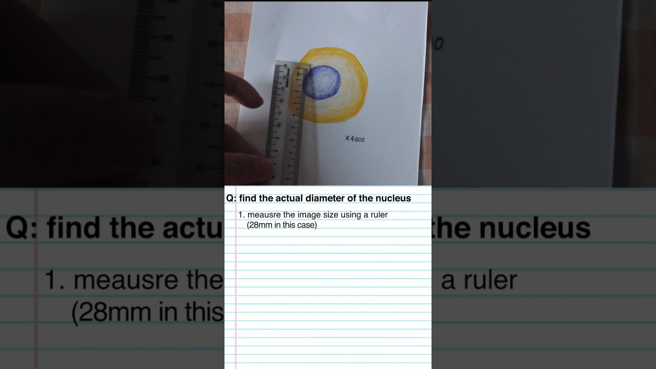
how to calculate actual diameter from a photomicrograph #alevelbiology #igcsebiology #alevelrevision
how to calculate actual diameter from a photomicrograph #alevelbiology #igcsebiology #alevelrevision
Related image with a detail of the photomicrograph shown in fig 1b where the arrows download scientific
 Detail Of The Photomicrograph Shown In Fig. 1B, Where The Arrows... | Download Scientific ...")


Related image with a detail of the photomicrograph shown in fig 1b where the arrows download scientific


About "A Detail Of The Photomicrograph Shown In Fig 1b Where The Arrows Download Scientific"
Comments are closed.