
Cervical Spine MRI: Normal Anatomy | E-Anatomy
Cervical Spine MRI: Normal Anatomy | E-Anatomy This module of human anatomy is useful for residents and students who wish to learn the basics of the anatomy of the cervical spine in mri on a 1.5 tesla device. This case illustrates the normal anatomy features found in the cervical spine mri. please refer on normal spine imaging examples article for more examples, including other modalities such as radiographs and ct.

Cervical Spine MRI: Normal Anatomy | E-Anatomy
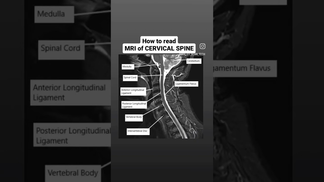
Cervical Spine MRI: Normal Anatomy | E-Anatomy This mri cervical spine axial anatomy tool is absolutely free to use. this section of the website will explain large and minute details of axial cervical spine cross sectional anatomy. This photo gallery presents the anatomical structures found on cervical spine mri (t2 weighted axial and sagittal views). This article illustrates normal spinal anatomy as defined by mr imaging, describes commonly used spinal mr imaging protocols, and discusses associated common artifacts. Welcome to today's lesson on the mri of the cervical spine. in this lesson, we will look at some anatomy on sagittal and axial scans. let's get started. starting with the sagittal view, slices start laterally. we can see the cerebellum located superiorly.

Cervical Spine MRI: Normal Anatomy | E-Anatomy
Cervical Spine MRI: Normal Anatomy | E-Anatomy This article illustrates normal spinal anatomy as defined by mr imaging, describes commonly used spinal mr imaging protocols, and discusses associated common artifacts. Welcome to today's lesson on the mri of the cervical spine. in this lesson, we will look at some anatomy on sagittal and axial scans. let's get started. starting with the sagittal view, slices start laterally. we can see the cerebellum located superiorly. High resolution surface coil mr imaging reveals intricate anatomic detail of the cervical spinal canal and its neurovascular contents. appreciation of the normal neurovascular anatomy provides a scientific foundation for the detection of disease. Understanding the anatomy revealed through a cervical spine mri is crucial for both healthcare professionals interpreting the scans and patients seeking clarity about their condition. In this section, the normal spinal anatomy is illustrated using both mri and myelography, as well as some more rarely used imaging techniques such as spinal angiography. mri cervical spine: investigation of choice for cervical myelopathy and radiculopathy. The human vertebral column or spine has five distinct anatomical regions: cervical, thoracic, lumbar, sacral, and coccygeal. however, the cervical spine is a potential area of importance due to its proximity to the head, containment of the upper spinal cord, and vertebral arteries that contribute to the posterior circulation of the brain. seven cervical vertebrae, combined with cartilages.

Cervical Spine MRI: Normal Anatomy | E-Anatomy
Cervical Spine MRI: Normal Anatomy | E-Anatomy High resolution surface coil mr imaging reveals intricate anatomic detail of the cervical spinal canal and its neurovascular contents. appreciation of the normal neurovascular anatomy provides a scientific foundation for the detection of disease. Understanding the anatomy revealed through a cervical spine mri is crucial for both healthcare professionals interpreting the scans and patients seeking clarity about their condition. In this section, the normal spinal anatomy is illustrated using both mri and myelography, as well as some more rarely used imaging techniques such as spinal angiography. mri cervical spine: investigation of choice for cervical myelopathy and radiculopathy. The human vertebral column or spine has five distinct anatomical regions: cervical, thoracic, lumbar, sacral, and coccygeal. however, the cervical spine is a potential area of importance due to its proximity to the head, containment of the upper spinal cord, and vertebral arteries that contribute to the posterior circulation of the brain. seven cervical vertebrae, combined with cartilages.

Cervical Spine MRI: Normal Anatomy | E-Anatomy
Cervical Spine MRI: Normal Anatomy | E-Anatomy In this section, the normal spinal anatomy is illustrated using both mri and myelography, as well as some more rarely used imaging techniques such as spinal angiography. mri cervical spine: investigation of choice for cervical myelopathy and radiculopathy. The human vertebral column or spine has five distinct anatomical regions: cervical, thoracic, lumbar, sacral, and coccygeal. however, the cervical spine is a potential area of importance due to its proximity to the head, containment of the upper spinal cord, and vertebral arteries that contribute to the posterior circulation of the brain. seven cervical vertebrae, combined with cartilages.
How to read MRI of Cervical Spine #viral #mri #shorts
How to read MRI of Cervical Spine #viral #mri #shorts
Related image with cervical spine mri normal anatomy e anatomy


Related image with cervical spine mri normal anatomy e anatomy


About "Cervical Spine Mri Normal Anatomy E Anatomy"
Comments are closed.