Flow Diagram Of The Preoperative Three Dimensional Reconstruction Download Scientific Diagram

Flow Diagram Of The Preoperative Three-dimensional Reconstruction... | Download Scientific Diagram
Flow Diagram Of The Preoperative Three-dimensional Reconstruction... | Download Scientific Diagram The aim of this retrospective study was to compare the use of 3d image reconstruction, and thin section multi detector computed tomography (mdct) in the preoperative evaluation of the. We compared three dimensional image reconstruction and thin section mdct images for preoperative evaluation of the sa branches of segmentectomy. our results indicate that the thin section mdct images provide a better evaluation of smaller sa branches.

Flow Diagram Of The Preoperative Three-dimensional Reconstruction... | Download Scientific Diagram
Flow Diagram Of The Preoperative Three-dimensional Reconstruction... | Download Scientific Diagram Therefore, this project aimed to develop an easy to use, low cost, and accurate workflow of generating medical 3d models. the workflow will help improving the medical education and career training of surgeons. Three dimensional (3d) reconstruction of human organs has gained attention in recent years due to advances in the internet and graphics processing units. in the coming years, most patient care will shift toward this new paradigm. We had a 3d printed model for preoperative planning, and surgery was performed using a transorbital approach to extract the wood 14 days after the accident. Through the analysis of the most recent works, we provide an overview of the workflows and tools that are currently used for 3d reconstruction from histologic sections and address points for future work, such as a missing common file format or computer aided analysis of the reconstructed model.

Flow Diagram Of Three-dimensional Reconstruction Method. | Download Scientific Diagram
Flow Diagram Of Three-dimensional Reconstruction Method. | Download Scientific Diagram We had a 3d printed model for preoperative planning, and surgery was performed using a transorbital approach to extract the wood 14 days after the accident. Through the analysis of the most recent works, we provide an overview of the workflows and tools that are currently used for 3d reconstruction from histologic sections and address points for future work, such as a missing common file format or computer aided analysis of the reconstructed model. Flow diagram depicting the design of the control study showing h3dr pathway and standard cta guided surgery. h3dr, hyper accuracy 3 dimensional reconstruction; cta, computed tomography angiography. This paper reviews critical aspects of 3d printing for preoperative planning and surgical training, starting with an overview of the process flow and 3d printing techniques, followed by their applications spanning across multiple organ systems in the human body. Flow chart for 3d reconstruction of medical images & applications the medical images (ct/mri images) are segmented into 2d information and integrated into 3d region growth. (a) preoperative cta 3 dimensional volume rendering reconstruction showing a proximally angulated neck, infrarenal aneurysmal neck, and abdominal aortic aneurysm sac. (b) intraoperative angiography showing the stiff guidewire status, herein called “kabedon.” tension was maintained through the guidewire along the large curvature of the aaa.

Preoperative Three‐dimensional Reconstruction Obtained From Computed... | Download Scientific ...
Preoperative Three‐dimensional Reconstruction Obtained From Computed... | Download Scientific ... Flow diagram depicting the design of the control study showing h3dr pathway and standard cta guided surgery. h3dr, hyper accuracy 3 dimensional reconstruction; cta, computed tomography angiography. This paper reviews critical aspects of 3d printing for preoperative planning and surgical training, starting with an overview of the process flow and 3d printing techniques, followed by their applications spanning across multiple organ systems in the human body. Flow chart for 3d reconstruction of medical images & applications the medical images (ct/mri images) are segmented into 2d information and integrated into 3d region growth. (a) preoperative cta 3 dimensional volume rendering reconstruction showing a proximally angulated neck, infrarenal aneurysmal neck, and abdominal aortic aneurysm sac. (b) intraoperative angiography showing the stiff guidewire status, herein called “kabedon.” tension was maintained through the guidewire along the large curvature of the aaa.

Three-dimensional-model Reconstruction Of Preoperative Computed... | Download Scientific Diagram
Three-dimensional-model Reconstruction Of Preoperative Computed... | Download Scientific Diagram Flow chart for 3d reconstruction of medical images & applications the medical images (ct/mri images) are segmented into 2d information and integrated into 3d region growth. (a) preoperative cta 3 dimensional volume rendering reconstruction showing a proximally angulated neck, infrarenal aneurysmal neck, and abdominal aortic aneurysm sac. (b) intraoperative angiography showing the stiff guidewire status, herein called “kabedon.” tension was maintained through the guidewire along the large curvature of the aaa.
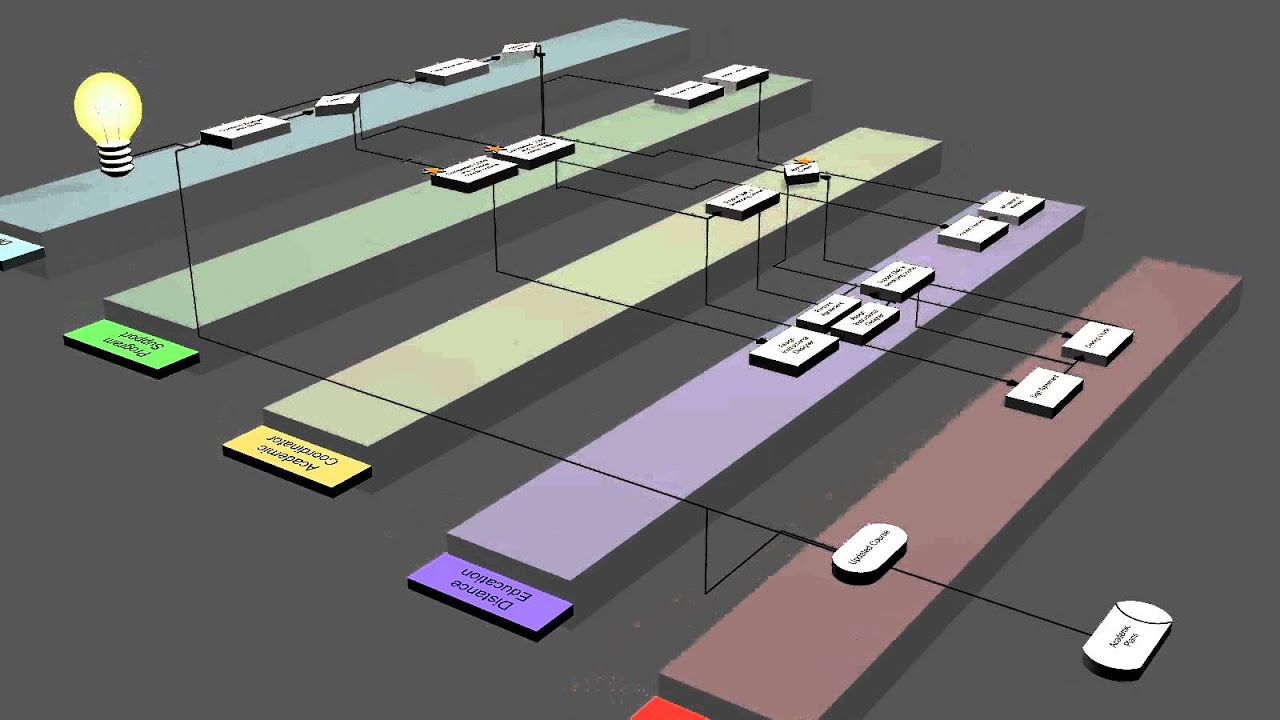
3D Process Flow - SwimLanes Animation
3D Process Flow - SwimLanes Animation
Related image with flow diagram of the preoperative three dimensional reconstruction download scientific diagram


Related image with flow diagram of the preoperative three dimensional reconstruction download scientific diagram


About "Flow Diagram Of The Preoperative Three Dimensional Reconstruction Download Scientific Diagram"
Comments are closed.