
-Photomicrograph Showing High | Download Scientific Diagram
-Photomicrograph Showing High | Download Scientific Diagram Download scientific diagram | photomicrograph showing from publication: antagonism of trichoderma viride and effects of extracted water soluble compounds from trichoderma species and. Learn about microscopy and drawing for your a level biology course. find information on microscopy, graticule use and scientific drawings.

Photomicrograph Showing | Download Scientific Diagram
Photomicrograph Showing | Download Scientific Diagram Photomicrograph showing: a) gram staining of tissue showing presence of gram negative slightly curved rods of vibrio spp. in between rbc's (arrows) (gram stainx1000). The evident scientific microscopy resource center gallery of phase contrast photomicrography contains a large collection of images taken under a wide variety of conditions using specimens ranging from fossilized dinosaur bones to soft tissues from human and plant pathology. Current study aimed at determining the consequence of radiofrequency (≈1800 mhz) electromagnetic radiation (rf emr) on the histological, hematological and histochemical properties of selected. This page outlines essential learning goals for interpreting microscopy results, focusing on identifying microscopy types based on scale, magnification, and resolution. it offers guidance for ….

Photomicrograph Illustrating | Download Scientific Diagram
Photomicrograph Illustrating | Download Scientific Diagram Current study aimed at determining the consequence of radiofrequency (≈1800 mhz) electromagnetic radiation (rf emr) on the histological, hematological and histochemical properties of selected. This page outlines essential learning goals for interpreting microscopy results, focusing on identifying microscopy types based on scale, magnification, and resolution. it offers guidance for …. This is the first study showing the biological potential of silver nanoparticles synthesized from an aqueous extract of p. oxalicum (poagnps). Peripheral blood smear (x100) of case 1, (a) photomicrograph showing red cells that are microcytic (m), hypochromic (h), or microcytic hypochromic (m/h). a small lymphocyte (l) is shown for. Photomicrograph of duodenum of negative control group (a) showing intact intestinal villi lined by simple columnar epithelium (ep), lamina propria (lp), and lamina muscularis (lm). Photomicrograph of a section of the lung from the control group showing the thin walled alveoli (a) separated by interalveolar septa (s). the alveoli are lined by squamous epithelium (e).
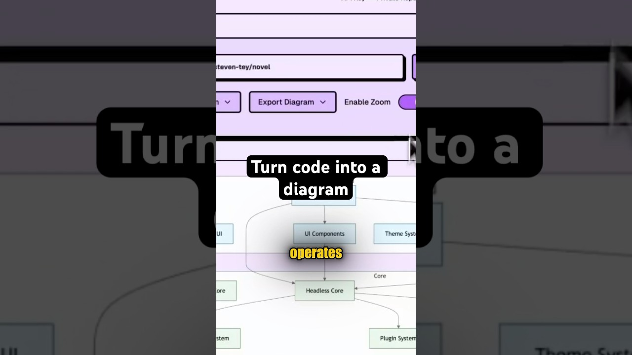
Turn any code into a diagram #creatorsearchinsights
Turn any code into a diagram #creatorsearchinsights
Related image with photomicrograph showing download scientific diagram


 Detail Of The Photomicrograph Shown In Fig. 1B, Where The Arrows... | Download Scientific ...")
Related image with photomicrograph showing download scientific diagram


About "Photomicrograph Showing Download Scientific Diagram"
Comments are closed.