
JCI Insight - Highly Multiplexed Imaging Reveals Prognostic Immune And Stromal Spatial ...
JCI Insight - Highly Multiplexed Imaging Reveals Prognostic Immune And Stromal Spatial ... The cytoviewer package offers a rich set of features for highly multiplexed imaging data visualization in r that seamlessly integrates with the workflow for image and single cell data analysis. Here, we explore the current state of the art computational pipelines for multiplexed tissue imaging, highlight achievements and challenges, and envision a future in which we integrate.

Visualization And Analysis Of Highly Multiplexed Imaging Data - YouTube
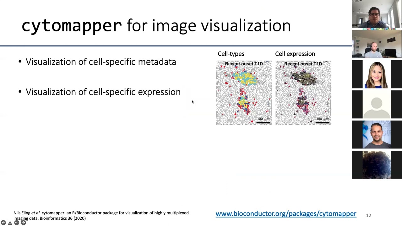
Visualization And Analysis Of Highly Multiplexed Imaging Data - YouTube Here, we describe cytomapper, a computational tool written in r, that enables visualization of pixel and cell level information obtained by multiplexed imaging. to illustrate its utility, we analysed 100 images obtained by imaging mass cytometry from a cohort of type 1 diabetes patients. Stitching microscope images into a mosaic is an essential step in the analysis and visualization of large biological specimens, particularly human and animal tissues. recent approaches to highly multiplexed imaging generate high plex data from sequential rounds of lower plex imaging. We introduce scimap, a python package specifically crafted to address these challenges. with scimap, users can efficiently preprocess, analyze, and visualize large datasets, facilitating the exploration of spatial relationships and their statistical significance. Here, we review the development and workflow of mci, show a complete mci data analysis workflow with a detailed introduction on each step, summarize the advanced bioinformatics tools and prospect the future direction of analytical strategies and technology application.

JCI Insight - Highly Multiplexed Imaging Reveals Prognostic Immune And Stromal Spatial ...
JCI Insight - Highly Multiplexed Imaging Reveals Prognostic Immune And Stromal Spatial ... We introduce scimap, a python package specifically crafted to address these challenges. with scimap, users can efficiently preprocess, analyze, and visualize large datasets, facilitating the exploration of spatial relationships and their statistical significance. Here, we review the development and workflow of mci, show a complete mci data analysis workflow with a detailed introduction on each step, summarize the advanced bioinformatics tools and prospect the future direction of analytical strategies and technology application. Here, we present an end to end workflow for multiplexed tissue image processing and analysis that integrates previously developed computational tools to enable these tasks in a user friendly. Viv addresses a critical limitation of most web based bioimaging viewers by removing a dependency on server side rendering, offering a flexible toolkit for browsing multi terabyte datasets on both mobile and desktop devices—without software installation. Overall, clustering local indicators of spatial association is an effective approach for quantifying high dimensional spatial ordering in highly multiplexed in situ imaging cytometry data. Summary: highly multiplexed imaging enables single cell resolved detection of numerous biological molecules in their spatial tissue context. interactive data visualization of multiplexed imaging data is necessary for quality control and hypothesis examination.

Highly Multiplexed Image Analysis Of Intestinal Tissue Sections In Patients With Inflammatory ...
Highly Multiplexed Image Analysis Of Intestinal Tissue Sections In Patients With Inflammatory ... Here, we present an end to end workflow for multiplexed tissue image processing and analysis that integrates previously developed computational tools to enable these tasks in a user friendly. Viv addresses a critical limitation of most web based bioimaging viewers by removing a dependency on server side rendering, offering a flexible toolkit for browsing multi terabyte datasets on both mobile and desktop devices—without software installation. Overall, clustering local indicators of spatial association is an effective approach for quantifying high dimensional spatial ordering in highly multiplexed in situ imaging cytometry data. Summary: highly multiplexed imaging enables single cell resolved detection of numerous biological molecules in their spatial tissue context. interactive data visualization of multiplexed imaging data is necessary for quality control and hypothesis examination.

VCG Harvard | Visinity: Visual Spatial Neighborhood Analysis For Multiplexed Tissue Imaging Data
VCG Harvard | Visinity: Visual Spatial Neighborhood Analysis For Multiplexed Tissue Imaging Data Overall, clustering local indicators of spatial association is an effective approach for quantifying high dimensional spatial ordering in highly multiplexed in situ imaging cytometry data. Summary: highly multiplexed imaging enables single cell resolved detection of numerous biological molecules in their spatial tissue context. interactive data visualization of multiplexed imaging data is necessary for quality control and hypothesis examination.

Panel Discussion: Clustering Analysis Of Highly Multiplexed Immunofluorescence Assays Analyzed ...
Panel Discussion: Clustering Analysis Of Highly Multiplexed Immunofluorescence Assays Analyzed ...
Visualization and Analysis of Highly Multiplexed Imaging Data
Visualization and Analysis of Highly Multiplexed Imaging Data
Related image with visualization and analysis of highly multiplexed imaging data


Related image with visualization and analysis of highly multiplexed imaging data


About "Visualization And Analysis Of Highly Multiplexed Imaging Data"
Comments are closed.